Histology & Slide Scanning
Automated whole-slide scanning with stitching
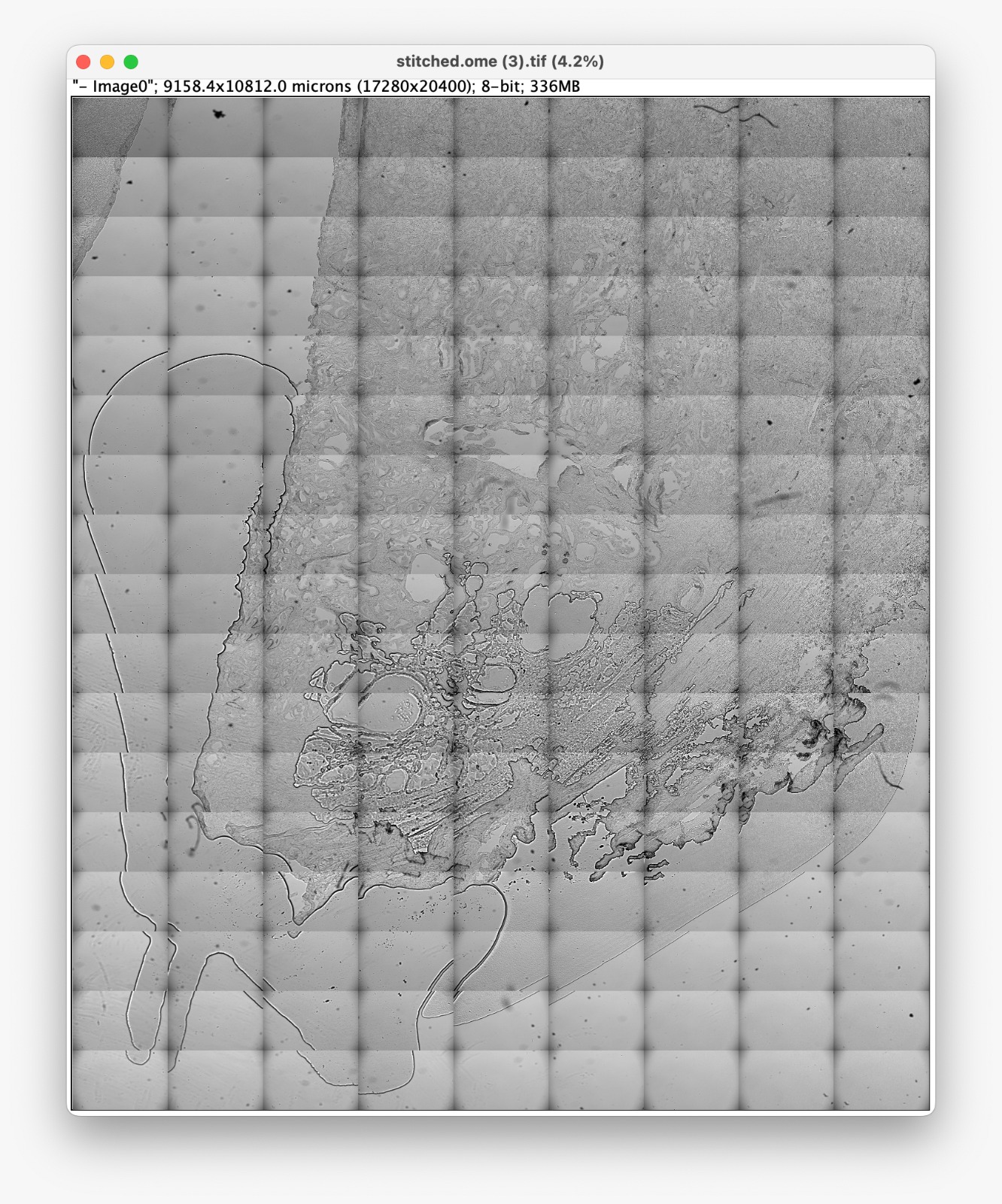
Turn your openUC2 FRAME into a digital pathology slide scanner. Automated XYZ stage movement captures overlapping tiles across the full 130x90 mm travel range, then stitches them into a seamless whole-slide image. Process H&E, IHC, or special stains at 10x-40x magnification with consistent Koehler illumination. Postprocessing will be available soon. Raw Images e.g. as OME.TIF or OME.Zarr or simply individual Tif images can be exported immediately.
Technical specifications
| Scan area | Up to 130 x 90 mm (full stage travel) |
|---|---|
| Resolution | 10x to 40x objectives |
| Illumination | Transmitted white LED, Koehler-capable |
| Stitching | Automated overlap and alignment in ImSwitch |
| Focus | Software autofocus per tile |
| Output | Tiled TIFF, OME-ZARR, or pyramidal formats |
Key capabilities
Full-slide coverage
Scan up to 130x90 mm — enough for standard glass slides and multiple sections.
Automated tile stitching
ImSwitch captures overlapping tiles and assembles them into a seamless image.
Software autofocus
Contrast-based autofocus runs per tile, maintaining sharpness across uneven sections.
Digital pathology output
Export in standard formats (OME-ZARR, pyramidal TIFF) for use in QuPath, OMERO, or custom viewers.
On the roadmap
Interested in histology & slide scanning?
Tell us about your imaging needs and we'll help you find the right configuration.